

Copyright 2015 © Nigerian Journal of Paediatrics. All Rights Reserved. . Powered by Pelrox Technologies Ltd
ISSN 03 02 4660 AN OFFICIAL JOURNAL OF THE PAEDIATRIC ASSOCIATION OF NIGERIA

|


Quick Navigation


Niger J Paediatr 2016; 43 (2):95 98
CASE
REPORT
Adedoyin OT
Acute glomerulonephritis
Afolayan FM
mimicking nephrotic syndrome
Buhari MO
Abdulkadir MB
Ibrahim OR
Akintade OO
Abdualzeez TA
DOI:http://dx.doi.org/10.4314/njp.v43i2.6
Accepted: 4th January 2016
Abstract :
Background:
Acute
(130/80mmHg and 150/100mmHg
post infectious glomerulonephritis
respectively) both systolic and
Adedoyin OT (
)
(APIGN) describes a wide range
diastolic blood pressure were
Afolayan FM, Abdulkadir MB
of
glomerulonephritis character-
greater than 99 percentile. Labora-
Ibrahim OR, Akintade OO
Abdualzeez TA
ized by an immunologic response
tory investigations revealed mas-
Department of Paediatrics and
of
the kidney to varieties of infec-
sive proteinuria of 4+ and 3+ re-
Child Health
tious agent commonly bacteria. It
spectively, haematuria, hypoalbu-
University of Ilorin Teaching Hospital,
is
characterized by an abrupt on-
minemia, and hyperlipidemia. The
Ilorin, Kwara State, Nigeria
set of haematuria, moderate
histology of
their renal tissues
Email: ooadedoyin@yahoo.com
oedema, hypertension and pro-
revealed features in keeping with
teinuria usually < 2g/dl.
How-
acute glomerulonephritis.
Buhari MO
ever, between 2013 and 2015, we
Conclusion:
Acute
glomeru-
Department of Pathology,
managed two children who had
lonephritis may present with fea-
University of Ilorin Teaching Hospital,
Ilorin, Kwara State, Nigeria.
2
histological diagnosis of post in-
tures of nephrotic syndrome in-
fectious glomerulonephritis but
cluding nephrotic range proteinu-
presented with full complement
ria. Hence in the event of the pres-
of
features of nephrotic syndrome
entation of a mixed feature of
including nephrotic range protein-
nephrotic-nephritis as obtained in
uria in addition to the features of
the case of these two children,
nephritis.
management should first be in the
Case reports: A
5year old
boy
line of AGN while awaiting the
and an 8 year old girl were admit-
renal hisotology outcome before
ted to our Paediatric Unit with
considering the commencement of
history of generalized body swell-
steroid.
ing, reduction in the volume of
urine and cough. There was no
Keywords: Acute
post infectious
antecedent sorethroat or skin rash.
glomerulonephritis, post strepto-
At
presentation, both patients had
coccal glomerulonephritis, massive
mild dyspnea, anasarca, massive
proteinuria.
ascitis, and hypertension
Introduction
remained an important public health problem.
3
The incidence of APIGN in the developing countries is
Acute post infectious glomerulonephritis (APIGN)
on
the increase but has however declined in the industri-
describes a wide range of glomerulonephritis character-
alized world. Globally, the incidence of acute PSGN
ized by an immunologic response of the kidney to varie-
was estimated at 472,000 cases per year, out of which
456,000 occurred in developing countries . The inci-
4
ties of infectious agent commonly bacteria
1,2,3
.It is char-
acterized by intra glomerular inflammation with cellular
dence of PSGN in the less developed world was re-
proliferation from an immunologic response to bacteria,
ported at 24.3 cases per 100,000 person/ year whereas in
viruses and protozoa. The most common form of
2
the industrialized countries, its estimated prevalence was
0.3 cases per 100,000 person/ year. Thus, it is a disease
5
APIGN is the post streptococcal glomerulonephritis
(PSGN) caused by nephritogenic strains of
group A
of
underdeveloped and developing countries commonly
beta hemolytic streptococcus
infection of
the throat
and
seen in children under the age of 15.
skin.
3,4
It
is an important cause of acute kidney injury in
children. Though, the true incidence of APIGN is not
The typical clinical presentation of PSGN which is a
known due to the frequency of subclinical form, it has
representative of a larger group of APIGN is an abrupt
96
onset of microscopic haematuria, oedema, hypertension,
showed massive proteinuria(4+), haematuria(3+). Serum
oliguria, azotemia, and proteinuria. The urinary excre-
4
creatinine was 57µmol/l. Lipid profile revealed hyper-
tion
of protein varies widely in PSGN but the rate is
cholesterolemia (7.1mmol/l) and hypertriglyceridaemia
generally less than 3g/ days which is
what is found in
(2.2mmol/l). Serum albumin(18g/l) and protein (20g/l)
nephrotic syndrome. The oedema in PSGN is due to
were markedly reduced.
retention of fluid and electrolyte (sodium). Unlike
4
There was no bacteria growth in both throat and urine
nephritic syndrome, nephrotic syndrome is characterized
culture. He was managed as a case of nephrotic-
by
massive proteinuria, hypoalbuminemia, hyperlipide-
nephritic syndrome and commenced on intravenous
mia and anasarca (generalized oedema).
frusemide, ceftriaxone, nifedipine. Blood pressure had
However, there can be an atypical presentation of
normalized by the fourth day on admission. There was
resolution of the edema by the 8 day on admission and
th
nephritic syndrome characterized by symptoms of both
nephritic and nephrotic syndrome. When this occurs,
by
day 12, proteinuria and haematuria were 1+ respec-
the glomerular lesion is at the mesangium and the mem-
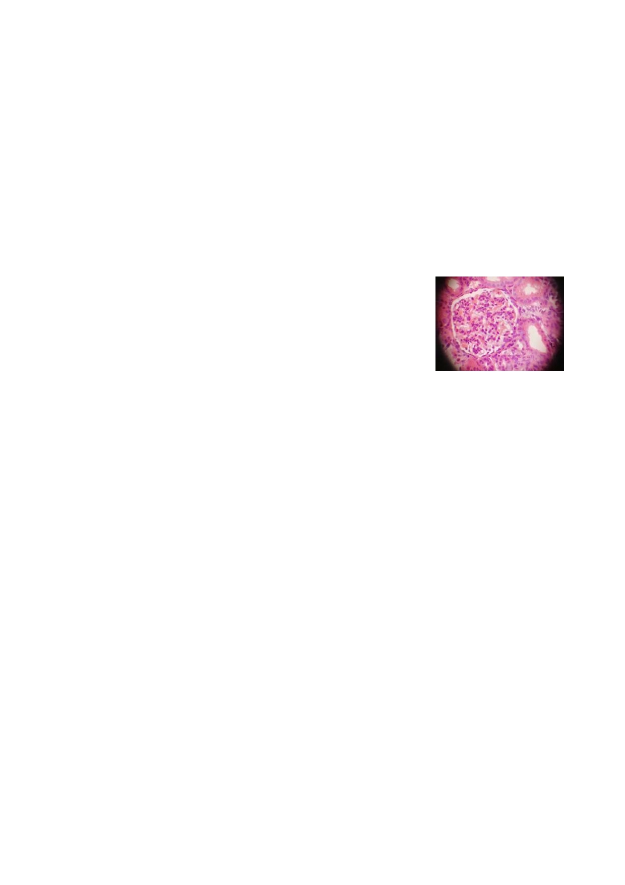
tively. Renal biopsy revealedendocapillary proliferation
brane. Patients with nephrotic range proteinuria in post
6
with infiltration of neutrophils and mesangial cellular
streptococcal glomerulonephritis has poor prognosis and
proliferation consistent with diffuse proliferative lesions
tend to develop hypertension and can also progress to
highly suggestive of post-infectious glomerulonephritis
chronic glomerulonephritis. We describe two children
(Figure 1).
who had histological diagnosis of post infectious glome-
rulonephritis but with full complement of features of
Fig 1: Endocapillary
prolif-
nephrotic syndrome including nephrotic range proteinu-
eration with infiltration of
ria in addition to features of nephritis.
neutrophils and mesangial
cellular proliferation consis-
tent with diffuse prolifera-
Case report
tive lesions highly sugges-
Case 1
tive of post-infectious
glomerulonephritis (×400).
A
5year old boy presented at the Emergency Paediatric
Unit with a six day history of generalized oedema and a
four day history of reduction in urinary output. There
Case 2
was progressive body swelling which initially started
from the face and was described as being worse in the
An
8year old girl presented with a seven day history of
morning but disappears as the day goes by which later
generalized body swelling, five day history of cough and
progressed to involve both legs and the abdomen and
a
three day history of difficulty with breathing. The
then became generalized. Four days after onset of body
swelling started from the face and progressed to involve
swelling, his parents noticed a reduction in urine pro-
the lower limb and abdomen. Swelling was worse early
duction in both volume and frequency. There was no
in
the morning and progressively resolved as the day
passage of coke colored or frothy urine and no prior
went by. There was no antecedent history of sore throat
history of a preceding sore throat or skin rash before
or
rash, though urine volume and frequency were re-
onset of symptoms. There was history of occasional
duced. There was no pain on micturition or passage of
cough but no difficulty in breathing or orthopnea. No
coke colored urine. No history of use of mercury con-
history of use of mercury containing soap, waddling in
taining soap, insect bite or ingestion of herbal remedies.
streams or recent blood transfusion. No family history of
Cough started two days after the onset of body swelling,
renal disease. Parents also attested to the use of herbal
was insidious in onset andnon paroxysmal. It was pro-
remedies. He was treated for malaria a month before
ductive of whitish sputum and not blood stained. Asso-
symptoms. There was no history of headache, convul-
ciated difficulty in breathing started a day after the onset
sion or loss of consciousness.
of
cough with worsening on lying supine. There was no
cyanosis
Physical examination revealed anasarca with platysma
lock and mild respiratory distress evidenced by flaring
Physical examination revealed anasarca and mild pallor
of
the alar nasi. He Weighed 21kg (expected 18kg),
(26%). She was in respiratory distress, respiratory rate;
height: 104cm, heart rate 84/min, respiratory rate:
42cpm with intercostal and subcostal recession. There
24cpm, blood pressure was 130/80 mmHg. There were
was a reduced breath sound with bilateral coarse basal
wide spread basal crepitations bilaterally, breath sounds
crepitations. Pulse rate; 122bpm, blood pressure was
were normal. There was no added heart sound. The ab-
150/100 mmHg. Abdomen was uniformly distended
domen was uniformly distended with flank fullness with
(girth-71 cm measured 15cm from the xiphisternum),
no
organ enlargement. Moderate ascitis was demonstra-
The liver was enlarged measuring 8cm below right cos-
ble by shifting dullness and the abdominal girth was
tal margin with a span of 16cm. There was massive as-
63.8cm measured 14cm from xiphisternal junction.
citis demonstrable by fluid thrill. There was no neuro-
There was also scrotal edema. There was no neurologi-
logical deficit.
cal deficit.
Initial laboratory test included complete blood counts
The complete blood counts revealed moderate anaemia
which reveal moderate anaemia(26%). Urinalysis
(PCV-33%)
with reversal of differential. Urinalysis
showed massive proteinuria (3+), haematuria (1+).

97
Serum creatinine was 392 µmol/l, urea was 19.1 mmol/l,
ria, hypercholesterolemia and hypoalbuminemia . How-
hypoalbuminemia (18g/l) , serum cholesterol was
ever, the presence of hypertension, haematuria and oe-
5.7mmol/l, serum triglyceride was 2.1 mmol/l. A diag-
dema which are the three most important triad of PSGN
nosis of nephrotic syndrome complicated by acute renal
makes it a mixed nephritic. The proteinuria and hema-
failure with pulmonary oedema was made.
turia of nephrotic syndrome is due to break in the
glomerular basement membranes, resulting from an in-
She was commenced on oxygen at 3Litre/ min, intrave-
flammatory response due to an immunologic mecha-
nous frusemide at 2mg/kg, then 1mg 8hrly. Intravenous
nism. Unlike nephritic syndrome, massive proteinuria
ceftriaxone was administered. She was given a bolus of
is
one of the most important findings in nephrotic
intravenous hydralazin followed by oral hydrochlorothi-
syndrome and it is due to damage to the podocyte within
azide, methyl dopa and captopril. By second day on
the glomerular membrane, leading to loss of negative
admission, respiratory distress had resolved, blood pres-
charge and thus increased permeability of the glomeruli.
sure was still elevated warranting another bolus of intra-
This leads to loss of albumin in urine, resulting in hypo-
venous hydralazin.
albuminemia cumulating into reduction in oncotic pres-
sure and finally oedema. Massive proteinuria is a rare
Blood
pressure
remained
between
140/110-
and atypical presentation in PSGN and when it occurs,
120/90mmHg in the first week on admission before a
it
is associated with severe disease and poor prognosis
significant improvement was made. She was maintained
In
PSGN, clinical recovery is the rule in 90% of cases.
on
the prescribed medication. She had progressive reso-
However with a finding of massive proteinuria, the
lution over the following 14 days into admission;
course of the microscopic proteinuria is likely to be pro-
oedema and ascitis resolved, weight declined, urinary
longed in atypical PSGN leading to chronic renal insuf-
output was within 1.1- 2.5ml/kg/hr and proteinuria and
ficiency. Thus early differentiation between a nephrotic
haematuria were 1+ and 2+ respectively by the 12 day.
th
-nephritic overlap and a typical nephritic syndrome us-
Renal biopsy done 10 days into admission revealed
ing a renal biopsy as a tiebreaker is important because
features in keeping with diffuse proliferative lesions
their management and prognosis are quite different.
highly suggestive of post-infectious glomerulonephritis
Though, renal biopsy is not often needed in PSGN,
(Figure 1). Child recovered and was discharged for fol-
except in atypical presentation.
low up in the clinic.
Most studies that described an overlap of nephrotic and
nephritic syndrome were in adult population with a dif-
ferent etiology other than group A beta hemolytic strep-
tococcus. Most importantly in resource poor countries
7
Discussion
where there are few facilities for renal biopsy, most pa-
The
presence of the combination of features of
tient with true PSGN who had atypical presentation
nephrotic and nephritic syndrome results in a clinical
tending towards a nephrotic syndrome might be treated
impression of nephrotic-nephritic syndrome. However,
as
nephrotic using steroid which is clearly contraindi-
in
view of the presence of nephrotic syndrome, it was
cated in PSGN.
instructive to carry out a renal biopsy to delineate the
histological type. Furthermore renal biopsy was necessi-
tated to differentiate between AGN which would not
require steroid and NS which requires the commence-
Conclusion
ment of steroid to classify whether it was steroid
responsive or resistant. In the two patients , while we
While the presence of a nephrotic picture in a PSGN is
were expecting a non- Minimal change NS such as focal
atypical,
clinicians should not commence steroid in
segmental glomerular sclerosis (FSGS),
meme-
such mixed state until a renal biopsy has been carried
branoproliferative glomerulonephritis (MPGN) e.t.c we
out, hence a nephrotic -nephritic picture makes renal
obtained a diffuse proliferative acute glomerulonephritis
biopsy mandatory. On the other hand in resource poor
most probably post infectious.
setting where renal biopsy may be difficult to procure,
such patients should be managed first as PSGN while
The presence of features of haematuria, hypertension
watching the trend and progress for relapse which would
and indeed azotaemia in children with NS portends a
further confirm nephrotic syndrome as recurrence is rare
poor prognosis as they will likely be non-MCNS. Hence
in
PSGN or complete recovery if it is PSGN. In addi-
extra care is deployed once nephrotic syndrome is
tion , follow up for the occurrence of persistent protein-
accompanied by features of nephritic syndrome.
uria, haematuria and hypertension will help prevent long
The concomitant clinical presentation of these two cases
term morbidity and mortality.
was that of nephrotic syndrome with massive proteinu-
References
2.
Stratta P, Musetti C, Barreca A,
3.
Kanjanabuch T, Kittikowit W,
1.
Moroni G, Ponticelli C. Acute post
Mazzucco G. New trends of an old
Eiam-Ong S. An update on acute
-infective glomerulonephritis.
disease: The acute post infectious
postinfectious glomerulonephritis
Recenti Prog. Med. 2003;94:395
glomerulonephritis
at the beginning of
worldwide. Nat
Rev Nephrol.
the
new millenium. J.
Nephrol.
398.
2014;27(M):229 239.
2009;5:259 269.

98
4.
Eison TM, Ault BH, Jones DP,
6.
Alper C. The Kidney. In: Kumar
7.
Velasquez TG, Barrios EJ, Botello
Chesney RW, Wyatt RJ. Post-
V,
Abbas A, Fausto N, Aster J,
CD.
Nephrotic syndrome with a
streptococcal acute glomeru-
editors. Robbins and Contrans
nephritic component associated
lonephritis in children: Clinical
Pathologic Basis of Disease .
Saun-
with toxoplasmosis in an immuno-
features and pathogenesis.
Pediatr.
ders Elsevier; 2012. p. 982 3.
competent young man. Colomb
Nephrol. 2011;26:165 180.
Med. 2012;43:226 229.
5.
Rodriguez-Iturbe B, Rodriguez-
Iturbe B, Musser JM, Musser JM.
The
current state of poststrepto-
coccal glomerulonephritis. J Am
Soc Nephrol. 2008;19:1855 64.